Last month we told you about the CMU Mobile Vet team visiting a 5-year-old female to diagnose a blocked nasal passage with a foreign object. On November 6, 2025, the CMU veterinarian participated in a surgical procedure to remove that foreign body. The case was first examined on October 29, 2025, when radiographic imaging revealed a solid, elongated object lodged in the right nasal passage. The surgical team at the elephant hospital, Lampang planned and successfully performed the operation to remove the foreign body, with the mobile clinic vet assisting in monitoring vital signs throughout the procedure. The surgery was successful, and a wooden stick approximately 50 cm in length was removed. The wound was then closed, and a postoperative care plan was implemented.
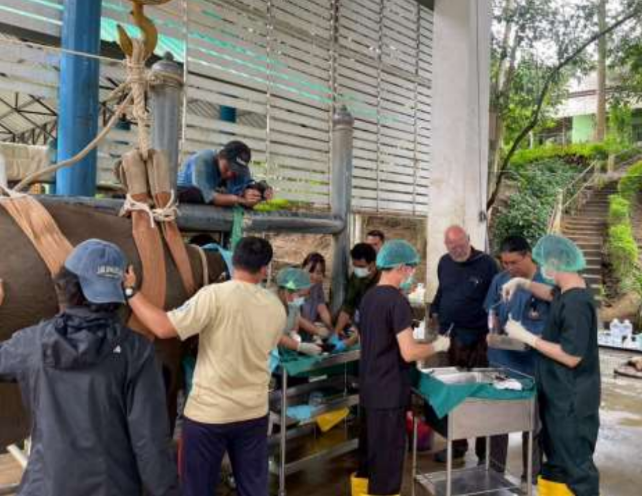
Top: Surgical procedure to remove the lodge object
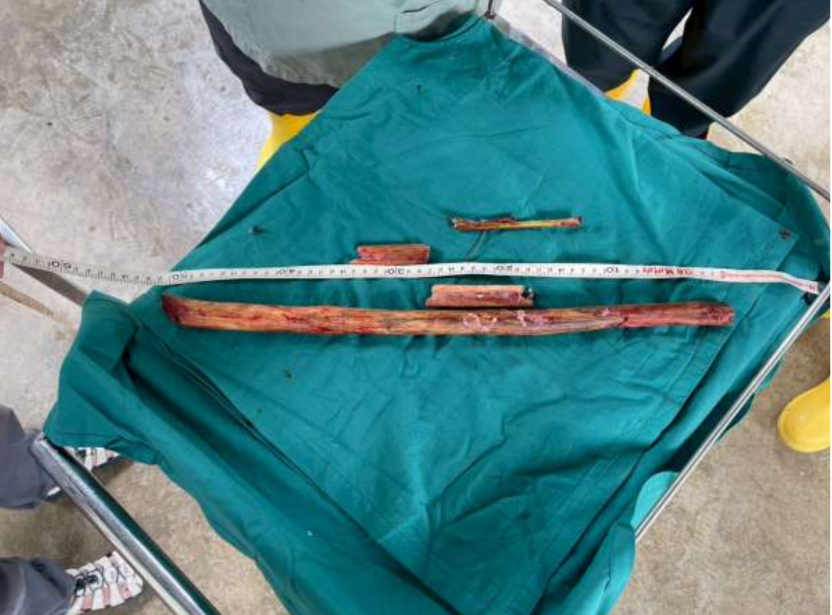
Below: The 50cm long stick removed from the youngsters trunk
At one camp, the owner reported that a 52-year-old male elephant had a wound on the sole of his foot. According to the history, the injury was likely caused by striking a tree stump, resulting in a laceration beneath the third and fourth nails of the right hind limb. The veterinarian examined the wound and found no active bleeding or signs of infection. Debridement was performed to allow the elephant to bear weight on the foot. The owner was instructed on proper wound care, advised to reduce the elephant’s walking, and to keep him secured in a dry and clean barn.
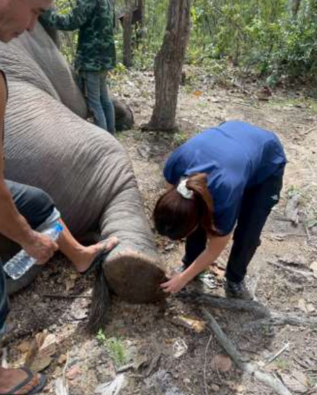
The veterinarian cleans and debris the wound on a rear pad
On the afternoon of November 11, the owner reported that a 3-year-7-month-old female elephant appeared lethargic and was spending more time lying down and resting than usual. Upon examination, the veterinarian obtained additional history and found that the calf was still eating and defecating normally. The mahout mentioned that the calf might not be getting enough rest due to disturbance from another young elephant and recent frequent heavy rain, which could have affected her sleeping pattern. Physical examination revealed no abnormalities—no petechiae or edema on the face, no fever, and no signs suggestive of infection. The veterinarian administered vitamins to boost immunity and advised improving the elephant’s resting environment. Blood samples were collected for health check and viral infection screening to further evaluate the elephant’s overall health condition.
A follow-up examination two days later revealed that the patient was bright and active with normal appetite, normal defecation, and no fever on rectal temperature. However, PCR testing was positive for EEHV, and antiviral therapy was initiated three times daily along with Vitamin C supplementation. Blood samples were collected for CBC, blood chemistry, and viral detection.
On a subsequent follow-up on November 17, the patient remained bright, alert and responsive with normal appetite, defecation, temperature and no petechiae or swelling. Antiviral medication and Vitamin C were continued, and blood was collected for monitoring. Thankfully, this time PCR results returned negative for EEHV, leading to discontinuation of Acyclovir while maintaining Vitamin C to support immune function.
Mouth check of the calf showing healthy tissue with no bruising or swelling
On 13 November 2025, the veterinarian conducted a health check on four calves. Upon examination, all calves appeared healthy, with no abnormalities detected. They showed normal growth for their age, ate and defecated normally, and displayed playful behavior typical of young elephants. Blood samples were collected from two calves that the mahouts were able to safely restrain, in order to assess immune status and perform viral detection. Encouragingly, the results showed no evidence of viral infection in either of the sampled elephants.
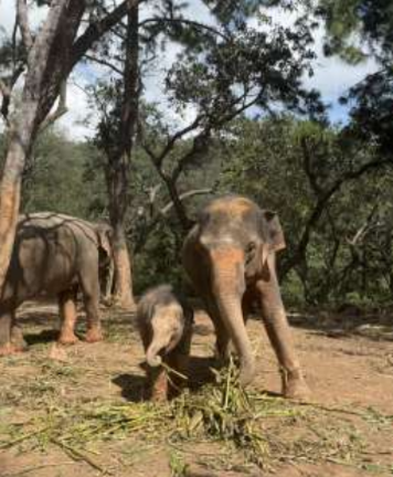
Happy healthy calf with mom after examination
Later in the month, the CMU team was called to check on two elephants who had been experiencing pruritus (itchy skin) and frequently rubbing their bodies against trees. History-taking and a physical examination revealed multiple skin lesions located behind the ears, on the legs, and on the hips, along with a heavy infestation of lice particularly noted behind the ears. An intramuscular anti-parasitic medication was administered, and the owner was advised to bathe the elephants thoroughly and brush the affected areas or apply an appropriate anti-louse treatment.
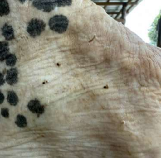
Back of the affected elephant’s ear showing the presence of lice
We are so grateful for the tireless efforts of the Chiang Mai University Mobile Veterinary Clinic team. It’s such a pleasure to read their reports when we get ones with such positive case outcomes. Help AES continue to support the efforts of the CMU vet team in caring for Thai elephants, by donating TODAY at www.asianelephantsupport.org/donate